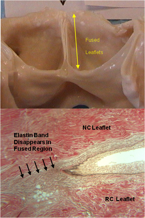
Adjacent valve leaflets fuse together along the commissures, accompanied by a loss of the laminar tissue structure.
Aortic Valve Fusion in LVAD Patients
PI: Karen May-Newman, Ph.D.
Aortic valve fusion is a pathological process in which fibrous tissue is deposited at the aortic valve commissures, creating adhesion between leaflets and preventing opening. Fusion, as well as aortic incompetence, has been associated with the implantation of LVADs, but no definitive link has been established between them.
Fusion was found in over 70% of the valves studied, and was often observed in multiple leaflets. Fusion length correlated loosely with the length of VAD support. Tissue from both fused and unfused valves showed unilateral fibrosis in the leaflets and loss of the laminar tissue structure that was related to the duration of VAD support. We believe that fusion is a remodeling response to the increased leaflet stress that occurs when left ventricular pressure is lowered by the LVAD. This biomechanical alteration increases the transvalvular pressure and compresses the tissue, reducing nutrient flow and the tissue's self-maintenance ability.
Recent Publications
- May-Newman K, Mendoza A, Abulon D, Joshi M, Kunda A, Dembitsky W. Geometry and fusion of aortic valves from pulsatile flow ventricular assist device patients. J Heart Valve Disease 20:149-158, 2011.
Return to Karen May-Newman's profile
SDSU Bioengineering Program 5500 Campanile Drive San Diego, CA 92182-1323
Engineering 326 Tel: (619) 594-6067 Fax: (619) 594-3599
